Visium v2 & Visium HD
Visium v2 and Visium HD for FFPE tissue are probe-based spatial transcriptomics solutions for high-resolution gene expression analysis. Both kits can be combined with protein staining enables whole-transcriptome profiling with spatially barcoded probes and limited protein co-detection, including a panel for 31 immune proteins. Visium HD offers enhanced spatial resolution for deeper insights into tissue architecture and cellular interactions.
Tissue: only FFPE
Species: Human & mouse tissue (custom probes available)
Capture: Probes targeting 18 transcripts
Resolution:
- Visium v2 → 55 µm spots
- Visium HD → 2 µm squares, continuous barcode carpet?
Cell Segmentation: Third-party BNF tools
ROI Sizes: 6.5 × 6.5 mm & 11 × 11 mm
Sequencing Depth: 125–400M reads/sample
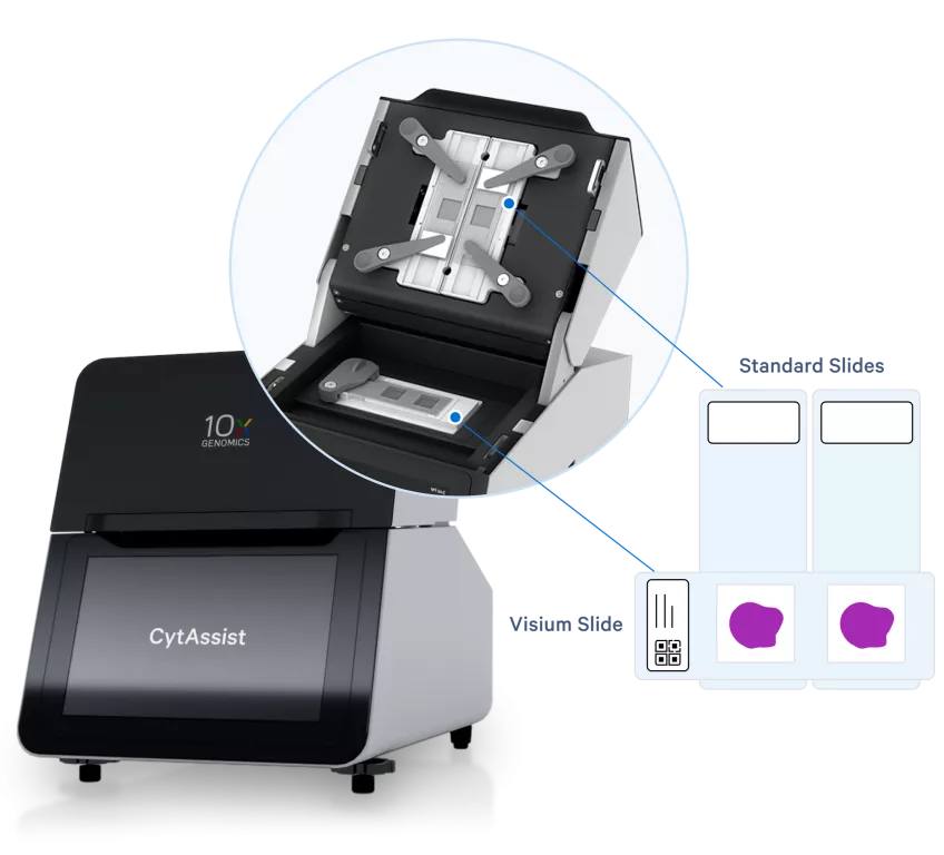
Links to 10x Genomics: Visium v2 and Visium HD
Sample Type: FFPE tissue
RNA Quality: DV200 >30% recommended for optimal data quality; ! For tissues with DV200 <30%, expect lower data yield and adjust expectations accordingly.
Tissue Morphology: Optional but recommended (DAPI/H&E staining) to assess suitability. Avoid artifacts like necrosis or cracks.
Protocols:
Slide Type: Coated/charged glass slides (see 10x Genomics recommendations)
Sections: one tissue section per slide
Thickness: 5 µm (range: 3–10 µm)
Visium area: 6.5 x 6.5 mm or 11 x11 mm (not for HD)
Labelling: Mark the area of interest to ensure correct preparation for CytAssist (ROI 6.5 x 6.5 mm or 11 x11 mm).
Replicates: Provide backup sections
Storage: Store in a desiccator for up to 6 months at room temperature
Protocols: FFPE Tissue Sectioning and Quality Assessment
Imaging: Before delivery, send images highlighting the region of interest (ROI) to guide RNA transfer selection. Basic H&E or DAPI, as well as IHC available
Waiting time for project start: 3 months
Lead times: 6-8 weeks
Request a consultation project service via iLab (link)
Our Process:
- Decrosslinking and Probe hybridization
- CytAssist Enabled Probe Release & Extension
- Library construction (QC)
- Pooling and Sequencing on NovaSeq X
- Data analysis using SpaceRanger software
- Data delivery through DDS